Unlocking the knee's secrets
Dive deep into the structure of the knee, understanding its vital components and how they contribute to stability and movement. This knowledge is crucial for comprehending ACL injuries and their impact.
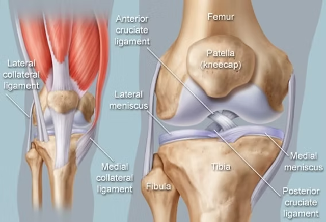
The Knee's Foundational Framework
The knee is a hinge joint, primarily formed by three major bones: the femur (thigh bone), the tibia (shin bone), and the patella (kneecap). Understanding their intricate connections is the first step in understanding knee function and vulnerability to injuries. 4 major ligaments maintain the stability of the knee. The anterior cruciate ligament (ACL), posterior cruciate ligament (PCL), medial cruciate ligament (MCL), and the lateral cruciate ligament (LCL). There are other key structures, such as the meniscus and cartilage, that act as shock absorbers and distribute the load across the joint evenly.
Ligaments versus Tendons: What's the difference?
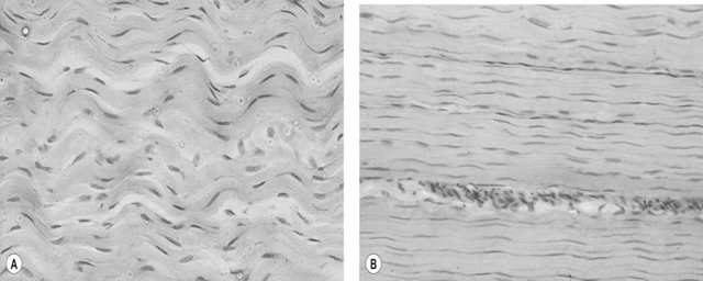
While often confused, ligaments and tendons play distinct roles. Ligaments connect bones to other bones, providing stability to joints, while tendons connect muscles to bones, facilitating movement. Both are crucial for knee function, but ligaments are directly involved in ACL injuries. Both of these structures are avascular, meaning that there is a lack of blood flow to them. The lack of blood flow means there is only limited healing that can occur, and often it is incomplete, and scar tissue develops, impacting overall functionality. Tendons and ligaments are both made primarily of collagen, but they differ in composition and organization. Tendons contain more tightly packed Type I collagen for strength and force transmission from muscle to bone, while ligaments have a slightly more varied collagen mix (including more Type III) and a looser arrangement, giving them greater flexibility to stabilize joints.
The Knee's Main Stabilizers: Four Crucial Ligaments
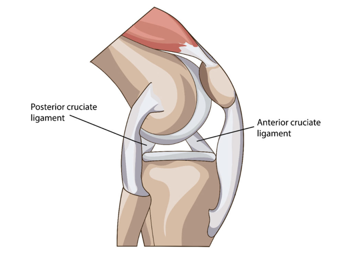
The ACL is the ligament that attaches the femur to the tibia in a diagonal fashion. The upper attachment sit the medial face of the lateral femoral condyle. The lower attachment sits on the tibia plateau. It is responsible for preventing dangerous anterior translation of the tibia. It is also a key contributor to rotational stability. it accounts for 85% of knee stability.
The PCL originates on the medial femoral condyle and then inserts into the posterior intercondylar are of the tibia. It is responsible for preventing the tibia from translating posteriorly past the femur.
The ACL and PCL form a cross in the center of the knee and because they are located inside the knee capsule there is very little blood flow compared to the LCL and MCL.
The MCL, or medial collateral ligament, is located on the inner side of the knee. It helps resist forces that push the knee inward, providing stability from side-to-side motion.
On the outer side is the LCL, or lateral collateral ligament. It stabilizes the knee against forces pushing outward and helps maintain overall lateral stability.
Understanding these components provides essential background knowledge on how the ACL works and why female athletes might be more prone to ACL injuries, considering anatomical, biomechanical, and hormonal differences (Hunter et al., 2021).
Create Your Own Website With Webador