Unraveling the Higher Risk in Women's Sports
Understanding why female athletes face a greater risk of ACL injuries is crucial. It's not one factor, but a combination of anatomical, hormonal, and biomechanical influences that all interact. Let's explore these interconnected elements.
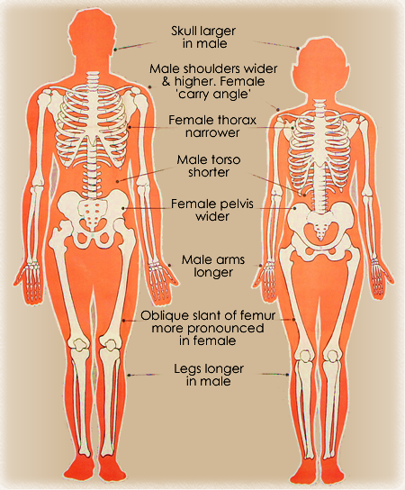
Anatomical differences
Female athletes are smaller and tend to have less muscle fibers to build upon. This means that women rely more heavily on the ligaments than the muscles, which also act as stabilizers to the knee. Women also have a smaller femoral notch which is where the ACL sits. . If that space is smaller, it can place additional mechanical stress on the ligament, especially during certain movements.
Female athletes often have a wider pelvis, leading to a larger Q-angle—the angle between the hip and the knee. This seemingly small difference significantly impacts how forces travel through the leg, creating more inward stress on the knee during movement. This increased inward motion places greater strain on the ACL, especially during high-intensity actions like cutting or landing.
Another factor is joint laxity. On average, women tend to have greater joint laxity than men. While some flexibility can be beneficial, too much laxity can reduce joint stability—meaning the knee may rely more heavily on ligaments like the ACL to maintain control.
Hormonal Influence
The ACL ligament itself has hormonal receptors which means this structure is directly impacted by hormonal changes in the body. Hormonal fluctuations, particularly those related to the menstrual cycle, can affect ligament laxity in female athletes. Estrogen and relaxin, for example, have been shown to increase joint looseness. This increased laxity can make the ACL more susceptible to injury, as the knee joint may have less inherent stability during dynamic movements.
Fluctuations in estrogen, progesterone, relaxin, and testosterone during the menstrual cycle can influence ligament strength and stability, particularly in the anterior cruciate ligament (ACL). Higher estrogen levels, especially around ovulation, are associated with decreased collagen stiffness and increased ligament laxity, which may make the ACL more vulnerable to injury. Progesterone and lower testosterone levels can further affect neuromuscular control and joint stability, collectively increasing the risk of ACL tears in certain phases of the cycle.
Understanding these cyclical changes is key to comprehensive injury prevention.
Visualize the Cycle of Hormones

Biomechanical Pattern Differences
Differences in biomechanical patterns, such as landing mechanics and cutting techniques, also contribute to higher ACL injury rates. Female athletes often exhibit tendencies such as knee valgus (knees caving inward) and a more upright landing posture, which can increase ACL load. These movement strategies, often subconscious, can be addressed through targeted training and neuromuscular control programs to reduce risk.
Another key factor is quadriceps dominance, a tendency for female athletes to rely more on the quadriceps than the hamstrings during movements like jumping or cutting. This imbalance is important because the quadriceps pull the tibia forward, increasing strain on the anterior cruciate ligament (ACL), while the hamstrings act as a protective force by pulling the tibia backward and stabilizing the knee. Ideally, these muscle groups should work in balance, but many female athletes demonstrate relatively lower hamstring strength and activation compared to their quadriceps. Research, including findings from the National Institutes of Health, suggests that women’s lower-body strength is often around 60–80% of men’s, which can contribute to reduced hamstring engagement. When the quadriceps dominate and the hamstrings are underutilized, the ACL must bear more of the load, increasing the risk of injury.
Another key factor is something called quadriceps dominance, which refers to a tendency for female athletes to rely more on the quadriceps than the hamstrings during movements like jumping or cutting. This imbalance is important because the quadriceps pull the tibia forward, increasing strain on the anterior cruciate ligament (ACL), while the hamstrings act as a protective force by pulling the tibia backward and stabilizing the knee. Ideally, these muscle groups should work in balance, but many female athletes demonstrate relatively lower hamstring strength and activation compared to their quadriceps. Research, including findings from the National Institutes of Health, suggests that women’s lower-body strength is often around 60–80% of men’s, which can contribute to reduced hamstring engagement. When the quadriceps dominate, and the hamstrings are underutilized, the ACL must bear more of the load, increasing the risk of injury.


Empowering prevention through knowledge
By understanding these complex interactions, we can develop more effective prevention strategies. We hope this information empowers you to advocate for and implement targeted interventions that protect female athletes. Explore our rehabilitation page for potential next steps.
Create Your Own Website With Webador